The incidence of neoplastic disease in the salivary glands is only 2 per 100.000 of the population, yet since the first introduction of fine needle aspiration biopsy (FNAB) these organs have provided an intriguing target.
FNAB is now a well established technique in the management of salivary gland disease but it also one of the most problematical tasks faced by cytopathologists.
Pleomorphic adenoma, is by far the most common of all salivary gland neoplasm constitute about 75% of major and minor salivary gland tumors. The great majority (78%) of pleomorphic adenomas are found in the superficial portion of the parotid glands, most of the tumors arise in the superficial portion of the parotid gland. The tumors occur most commonly in patients aged 30-50 years and are slightly more common in females.
The tumors are composed of epithelial and mioepithelial elements and a sparse to abundant stroma. The variability in histologic pattern is typically reflected in FNAB.
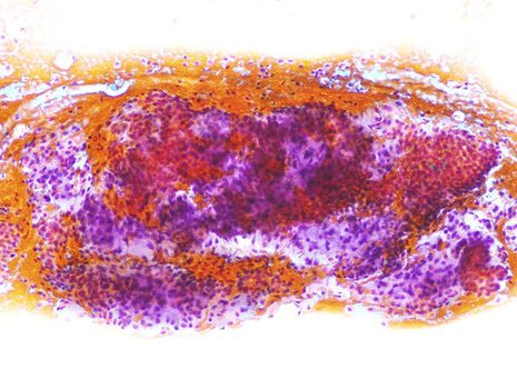
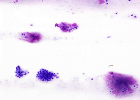
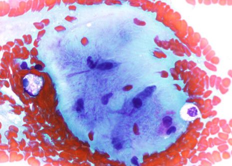
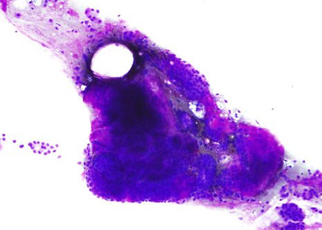
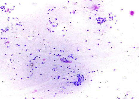
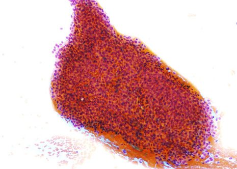
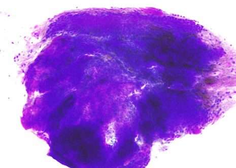
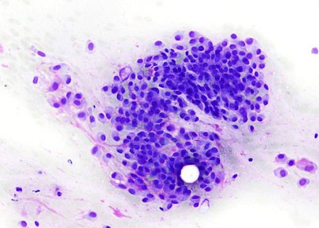
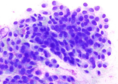
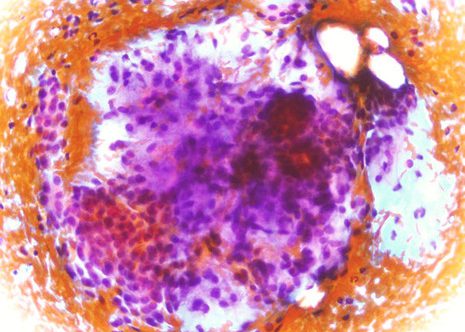
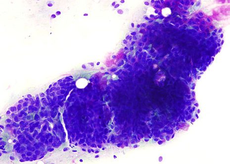
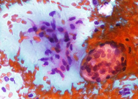
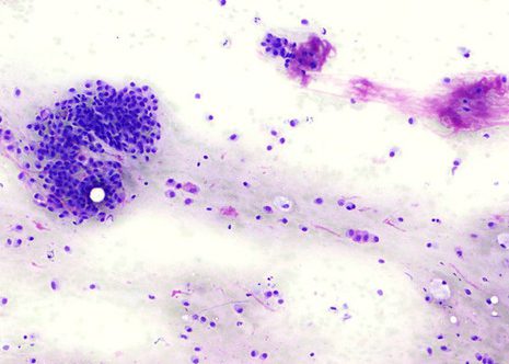
Pleomorphic adenoma is the lesion most likely to be encountered in FNAB practice. Aspirates contain plentiful epithelial cells which are closely intermingled with loose dusters of mesenchymal cells and fibrillary mucomyxomatous background substance. The cytologic features of pleomorphic adenoma are usually quite characteristic and the correct cytologic diagnosis can almost be established. The cytological diagnosis of the great majority of pleomorphic adenomas is quite straightforward.
The images correspond to the FNAB of a woman aged 39 years with a palpable left parotid nodular elevation. In this case the presence of characteristic cytological pattern should allow the diagnostic of pleomorphic adenoma. The distinction of pleomorphic adenoma from well differentiated adenoid cystic carcinoma is not always easy.
