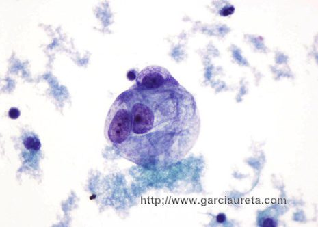
Cytologic examination of fluid obtained from the serous cavities is among the most common tasks performed in the practice of cytopathology. The diagnostic utility of effusion cytology is now well established making it routine when trying to find the cause of fluid accumulation in serous cavity.
Any neoplasm may involve a serous cavity and manifest as a malignant effusion. By for the commonest type of tumor to produce metastasis in the serous cavities is the broad group of adenocarcinomas most of them from the breast lung ovary and gastrointestinal tract.
Many effusions associated with cancer are a result of some form of indirect mechanism, such as a transudate that develops as a result of venous obstruction caused by neoplasm or an exudates that develops as a result of inflammatory changes in an organ secondary to the presence of neoplasm. These are extremely common situations and is such effusions neoplastic cells are not expected to be found. Even when a serous fluid contains neoplastic cells the number of such cells may vary considerably.
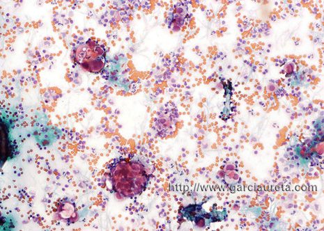
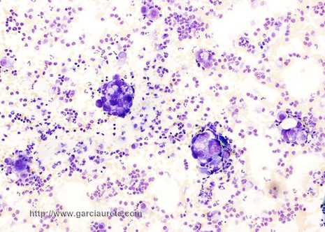
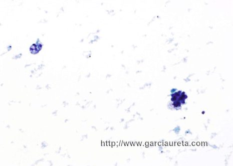
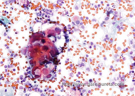
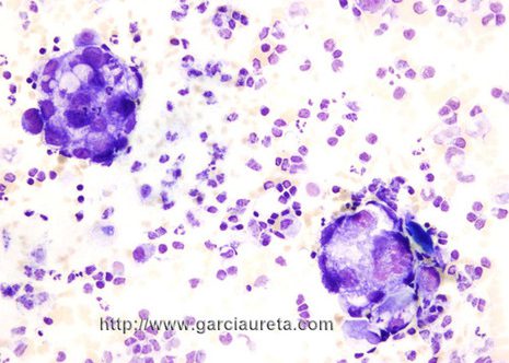
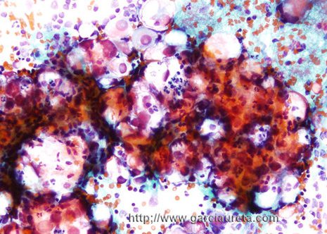
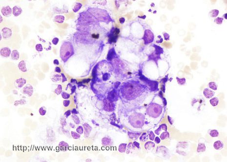
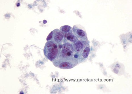
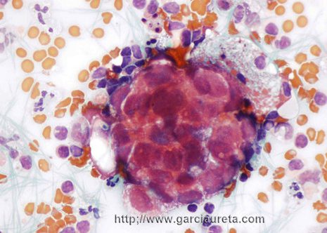
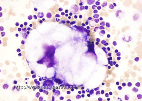
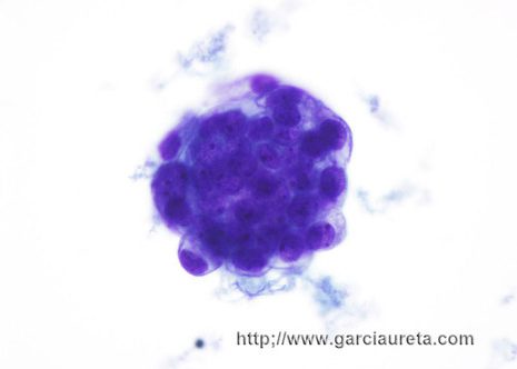
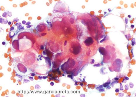
The cells may show the classic features of adenocarcinoma: a tendency to form smoothly contoured cohesive groups composed of large cells with eccentric malignant-appearing nuclei prominent nucleoli and vacuolated cytoplasm.
In theory identifying malignant cells in effusions should be a relatively easy task. Once one identifies the native mesothelial cells, the presence of a second population of neoplastic cells should permit a diagnosis of malignancy. In actual practice however it is a very difficult task because the many morphologic variations of mesothelial cells mimic many things.
